
Screening Mammography Redefined

By John F. Arnold, MD, MBA
The goal of any breast screening program is to find cancer early and reduce false positives. The American College of Radiology Commission on Breast Imaging has performed a thorough review of the evidence that annual mammography screening starting at age 40 for women of average risk can decrease breast cancer mortality by about 40%. Benefits of screening also include tumors being found earlier and the ability to discover high-risk lesions earlier.
 Supported by Research
Over 70% of cancers occur in dense breasts. Mammography has limitations in imaging dense breasts. The ASTOUND study is defined as adjunct screening with tomosynthesis or ultrasound in women with mammography negative dense breasts. This was a multicenter screening trial of tomosynthesis and adjunct ultrasound screening in women with dense breasts. Adjunct ultrasounds led to significantly higher incremental breast cancer detection, the rate per 1,000 screening mammograms was 7.1/1,000 screening exams for ultrasound and 4.0/1,000 screening exams for tomosynthesis. Three studies—European Asymptomatic Screening Study, SOMO•INSIGHT Clinical Study, and performance studies by Shane, et al—included adding 3-D automated whole breast ultrasound to screening mammography to improve the performance of mammographic interpretation. Adding 3-D automated whole breast ultrasound has shown a significant increase in invasive cancer detection with a nominal insignificant decrease in specificity. Eighty-five percent of cancers found with screening ultrasound are node negative invasive cancer.
OutPatient Imaging is Leading The Way
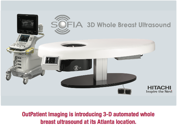
OutPatient Imaging is introducing 3-D automated whole breast ultrasound at its Atlanta location. Even better, OutPatient Imaging will be performing an automated 3-D screening breast ultrasound as an adjunct to all screening mammograms with no additional cost to the patient during the introductory phase. Ultrasound is fully automated and takes approximately 30 seconds per breast. The exam requires no external compression other than the patient’s own body weight. The 3-D images generated from the ultrasound will be interpreted by the radiologist as an adjunct to the patient’s screening mammogram. Three-D automated breast ultrasound does not utilize ionizing radiation as tomosynthesis uses, therefore it can reduce the radiation exposure to the patient.
OutPatient Imaging is proud of its commitment to women’s health and will continue to offer patients a choice for all of their imaging needs.
OutPatient Imaging • 2284 Peachtree Road N.W., Atlanta, GA 30309 • (404) 225-5674 • outpatientimaging.net
